PRODUCTS

Hepatica

Improving accuracy of liver health and volume assessment to support better pre-surgical decision making for patients with liver cancer.

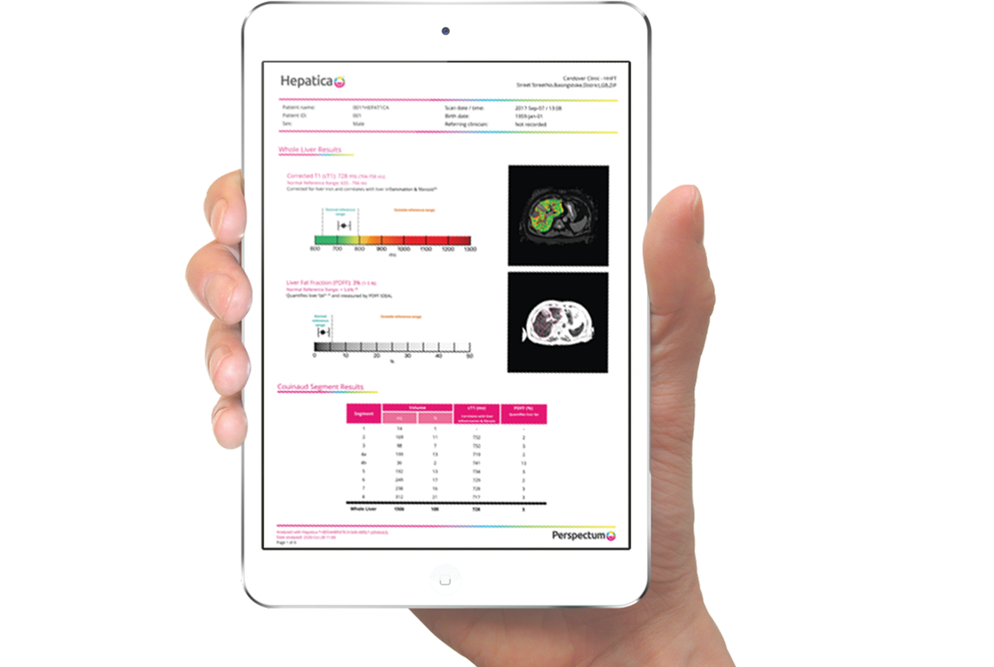
Liver health assessment and visualization on a single platform

  • Patient-friendly reports
  • Seamless integration into workflow through a PACS-integrated cloud-based service
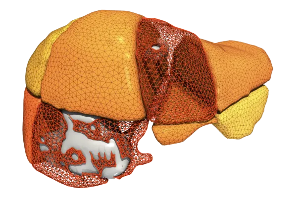
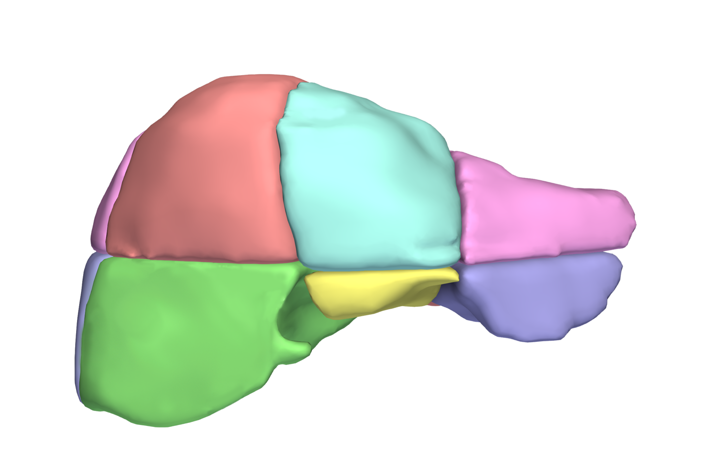
  • AI-driven delineation of liver volume and individual Couinaud segments
  • MRI-based, therefore no radiation risk, giving your patients a safer experience


Key Features

Stratify Risk

Supports identification of patients at risk of poor post-operative outcomes to inform surgical decision-making.(2)

Improve Outcomes

Clinically validated metrics to improve the accuracy of pre-operative liver health assessment.(1-5)

Accurate

Accurate and robust quantifiable metrics, which can assist in better estimation of future liver remnant volume and health calculations.(1)

Informs clinical decisions

Quantitative assessment of an individual’s liver health and volume presented in one comprehensive report.

What is Hepatica?

  • Hepatica is a clinical and surgical decision support tool.
  • Artificial intelligence-driven delineation of the liver and individual Couinaud segments in a single report containing quantitative metrics and images.
  • Demonstrated utility in liver cancer by identifying patients at risk of poor outcomes and longer hospital stay after resection surgery.2
  • Potential to enable cost savings through lowering the post-surgical complication rate, reducing the associated in-patient hospital stay and thus improving post-operative patient outcomes.


Couinaud segment analysis

Liver fibro-inflammation (cT1), fat (PDFF) and volume are quantified for individual Couinaud segments. This can assist clinicians in accurately predicting the future liver remnant (FLR) to inform surgical decision making.

Whole liver analysis:

Built on LiverMultiScan technology that provides validated biomarkers of liver health (cT1 and PDFF) across the whole liver and has been shown to predict clinical outcomes4–6. Provides 3D visualization and quantification of whole liver volume.

Supporting clinical decision making:

A study illustrating that pre-treatment assessment could improve post-treatment outcomes included two patients with similar pre-operative characteristics who had liver resection for colorectal liver metastases (CRLM) but different post-operative outcomes (Figure 1).3

Figure 1:  Quantitative multiparametric MRI allows safe surgical planning in patients undergoing liver resection for colorectal liver metastases: Report of two patients.3

Patient 1

  • Hepatica indicated high fibro-inflammation (cT1) and fat (PDFF)
  • Future liver remnant volume: 23%
  • Post-hepatectomy liver failure (PHLF)
  • Information may have changed surgical plan, averting PHLF that delayed the patient’s recovery

Patient 2

  • Hepatica indicated normal fibro-inflammation (cT1) and fat (PDFF)
  • Future liver remnant volume: 29%
  • Uneventful post-operative course and short hospital stay
  • Information would justify surgical plan


References

1. Mojtahed, A., Núñez, L., Connell, J., Fichera, A., Nicholls, R., Barone, A., Marieiro, M., Puddu, A., Arya, Z., Ferreira, C., Ridgway, G., Kelly, M., Lamb, H. J., Caseiro-Alves, F., Brady, J. M., & Banerjee, R. (2021.) Repeatability and reproducibility of deep-learning-based liver volume and Couinaud segment volume measurement tool. Abdominal Radiology (New York).

2. Mole, D. J., Fallowfield, J. A., Sherif, A. E., Kendall, T., Semple, S., Kelly, M., Ridgway, G., Connell, J. J., McGonigle, J., Banerjee, R., Brady, J. M., Zheng, X., Hughes, M., Neyton, L., McClintock, J., Tucker, G., Nailon, H., Patel, D., Wackett, A., … HepaT1ca Study Group. (2020.) Quantitative magnetic resonance imaging predicts individual future liver performance after liver resection for cancer. PloS One, 1512, e0238568.

3. Sethi, P., Thavanesan, N., Welsh, F. K., Connell, J., Pickles, E., Kelly, M., Fallowfield, J. A., Kendall, T. J., Mole, D. J., & Rees, M. 2021. Quantitative multiparametric MRI allows safe surgical planning in patients undergoing liver resection for colorectal liver metastases: Report of two patients. BJR Case Reports, 73, 20200172.

4. Dennis, A., Kelly, M. D., Fernandes, C., Mouchti, S., Fallowfield, J. A., Hirschfield, G., Pavlides, M., Harrison, S., Chakravarthy, M. V., Banerjee, R., & Sanyal, A. (2021.) Correlations Between MRI Biomarkers PDFF and cT1 With Histopathological Features of Non-Alcoholic Steatohepatitis. Frontiers in Endocrinology, 11, 1053.

5. Andersson, A., Kelly, M., Imajo, K., Nakajima, A., Fallowfield, J. A., Hirschfield, G., Pavlides, M., Sanyal, A. J., Noureddin, M., Banerjee, R., Dennis, A., & Harrison, S. (2021.) Clinical utility of MRI biomarkers for identifying NASH patients’ high risk of progression: A multi-center pooled data and meta-analysis. Clinical Gastroenterology and Hepatology: The Official Clinical Practice Journal of the American Gastroenterological Association, S1542-35652101056-9.

6. Jayaswal, A. N. A., Levick, C., Selvaraj, E. A., Dennis, A., Booth, J. C., Collier, J., Cobbold, J., Tunnicliffe, E. M., Kelly, M., Barnes, E., Neubauer, S., Banerjee, R., & Pavlides, M. (2020.) Prognostic value of multiparametric magnetic resonance imaging, transient elastography and blood-based fibrosis markers in patients with chronic liver disease. Liver International: Official Journal of the International Association for the Study of the Liver, 4012, 3071–3082.